The double helix: the structure of DNA
Before April 1953 no one knew how living things store and copy their hereditary information; after it, heredity could be described as a sequence written in a molecule, the ground on which genetics, molecular medicine and genome sequencing were built.
- Dated
- Place
- Cambridge, United Kingdom · Two laboratories in England: the Cavendish Laboratory in Cambridge and the Medical Research Council unit at King's College London
- Coordinates
- 52° 12′ N · 0° 07′ E (approximate)
- Remains
- Reconstructed
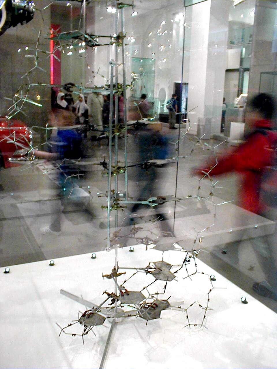
Alkivar, domaine public, via Wikimedia CommonsPDMOriginal record
Data saver is on. Images load only when you ask. You can change this in Settings at any time.
From the archivist
Four letters, A, T, G and C, and a rule: A faces T, G faces C. That is the whole alphabet.
In the spring of 1953, in a laboratory in Cambridge, two men took plates of metal cut in the workshop, one for each base, and clamped them on rods around a central pole, turning each rung a little on the one below, until the whole thing twisted upward like a spiral staircase and every piece fitted.
They had not made the measurements that told them it must be so. Those came from London, from X-ray photographs of DNA fibres taken by Rosalind Franklin and her student Raymond Gosling, and from the work of Maurice Wilkins. One image, a dark cross on a pale field, was shown to Watson without Franklin knowing.
Nature printed the three papers side by side. One sentence in the first said, with care, that the pairing suggested how life copies itself. It did. The original model was later taken apart; what the museum shows was rebuilt, around a few of the first plates.
Established
- On 25 April 1953 the journal Nature published three papers together: James Watson and Francis Crick's proposal of a double helix for DNA, a paper by Maurice Wilkins, Alexander Stokes and Herbert Wilson, and one by Rosalind Franklin and Raymond Gosling (Nature).
- Watson and Crick proposed two helical chains wound around the same axis, held together by pairs of bases, adenine with thymine and guanine with cytosine, and noted that this specific pairing 'immediately suggests a possible copying mechanism for the genetic material' (Watson and Crick 1953).
- The X-ray diffraction image later known as Photograph 51 was made in May 1952 at King's College London by Gosling, a doctoral student working with Franklin (Franklin and Gosling 1953; Cobb and Comfort 2023).
- Early in 1953 Wilkins showed that image to Watson, and Crick saw King's results summarised in a Medical Research Council report, without Franklin being asked (Cobb and Comfort 2023).
- Watson, Crick and Wilkins received the Nobel Prize in Physiology or Medicine in 1962; Franklin had died of cancer in 1958, at 37, and the prize is not awarded posthumously (Nobel Prize).
Interpretation
How much the Cambridge model owed to Franklin's data, and how fairly she was treated, has been argued for decades, often in the terms of Watson's own account of 1968. Historians Matthew Cobb and Nathaniel Comfort argued in 2023, from letters and an unpublished news article of 1953, that she was an equal member of a quartet that solved the structure rather than simply a victim whose photograph was taken. The model in the museum is a reconstruction; only some of its plates are original.
In 10,000 years
In 10,000 years, will those who read our genomes remember the four people who first saw how the molecule was folded?
Sources (6)
- Watson, J. D., Crick, F. H. C. Molecular Structure of Nucleic Acids: A Structure for Deoxyribose Nucleic Acid. Nature 171, 25 April 1953, p. 737-738. · doi.org
- Franklin, R. E., Gosling, R. G. Molecular Configuration in Sodium Thymonucleate. Nature 171, 25 April 1953, p. 740-741. · doi.org
- Wilkins, M. H. F., Stokes, A. R., Wilson, H. R. Molecular Structure of Deoxypentose Nucleic Acids. Nature 171, 25 April 1953, p. 738-740. · doi.org
- Cobb, M., Comfort, N. What Rosalind Franklin truly contributed to the discovery of DNA's structure. Nature 616, 2023, p. 657-660. · doi.org
- Nobel Prize Outreach. The Nobel Prize in Physiology or Medicine 1962. · www.nobelprize.org
- Science Museum Group Collection. Crick and Watson's DNA molecular model, object 1977-310. · collection.sciencemuseumgroup.org.uk